In our modern world, plastic is everywhere—from food packaging and water bottles to cosmetics and household items. But beneath its convenience lies a growing concern: the toxic impact of plastic chemicals on pregnant women and their unborn children. Mounting scientific evidence reveals that exposure to certain compounds in plastics can disrupt hormonal systems, impair fetal development, and increase the risk of serious health complications.
At the heart of this danger are chemicals like phthalates, bisphenols (such as BPA), and benzophenones, which are commonly added to plastics to enhance flexibility, durability, and UV resistance. These substances are known endocrine disruptors, meaning they interfere with the body’s hormonal balance. For pregnant women, whose hormone systems are already in overdrive to support fetal growth, this interference can be particularly harmful.
Phthalates, often referred to as “everywhere chemicals,” are especially concerning. They leach from plastic containers into food and beverages, are absorbed through the skin from personal care products, and even contaminate indoor air and dust. Once inside the body, phthalates can cross the placenta, exposing the developing fetus to their effects. Studies have linked prenatal phthalate exposure to increased risks of preterm birth, low birth weight, and developmental disorders such as ADHD and asthma.
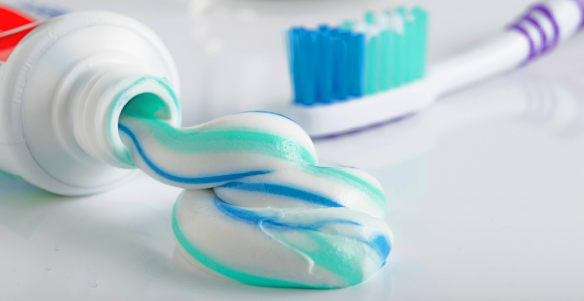
Bisphenols, including BPA, are another group of chemicals with alarming implications. Found in plastic bottles, food can linings, and thermal paper receipts, bisphenols mimic estrogen and can disrupt the delicate hormonal signaling crucial for fetal brain and organ development. Research suggests that exposure to BPA during pregnancy may be associated with behavioral problems, altered brain structure, and reproductive issues later in life.
The danger doesn’t stop with the products themselves. The entire lifecycle of plastic—from fossil fuel extraction to manufacturing and disposal—releases toxic byproducts like dioxins, which further contribute to hormonal disruption and immune system damage. These pollutants disproportionately affect vulnerable populations, including pregnant women and children, whose bodies are more sensitive to chemical interference.
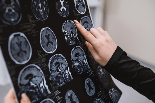
What makes these chemicals particularly insidious is their ubiquity and persistence. Unlike some toxins that degrade over time, many plastic-related compounds linger in the environment and accumulate in human tissues. The placenta, once thought to shield the fetus from harm, offers little protection against these invaders. As Boston College pediatrician Philip Landrigan warns, “The placenta provides no protection at all”.
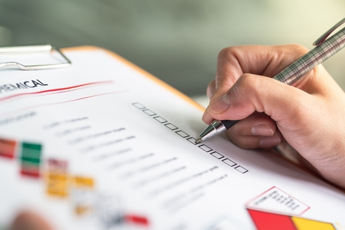
Despite the grim outlook, there are steps expectant mothers can take to reduce exposure. Avoiding plastic food containers, especially when heating food, choosing glass or stainless steel alternatives, and scrutinizing ingredient labels on personal care products can make a meaningful difference. Advocacy for stronger regulations and corporate accountability is also essential to protect future generations from the silent threat of plastic toxicity.
In a world saturated with synthetic materials, awareness is the first line of defense. By understanding the risks and making informed choices, pregnant women can help safeguard their health—and the health of their unborn children—from the hidden dangers lurking in plastic.